Suppose you are a doctor, on a long day shift, and reviewing hundreds of medical images. Fatigue sets in, and it becomes harder to focus, which could potentially affect diagnosis accuracy. This is where computer-aided diagnosis (CAD) steps in, enhancing the precision of diagnostics and supporting healthcare professionals in identifying diseases early. With the growing adoption of AI and machine learning, CAD is shaping the future of medical diagnostics by assisting radiologists and clinicians in detection and diagnosis tasks.
What Computer-Aided Detection and Diagnosis Can Propose
The use of CAD systems in clinical practice is generating excitement for several important reasons:
Increased Detection Accuracy: Computer-aided diagnosis systems are designed to assist radiologists in the detection of abnormalities, such as breast cancer, lung nodules, and liver lesions. Studies show that CAD improves the detection rate and helps reduce false negatives, enabling earlier detection of diseases.
Fatigue Reduction for Radiologists: Radiologists review countless imaging modalities daily. CAD systems reduce cognitive load by automating routine tasks, allowing healthcare professionals to focus on more complex diagnoses.
Faster Diagnosis: With CAD systems, medical imaging analysis is faster. Computer-aided detection enhances the speed at which images are processed, enabling radiologists to provide more timely diagnoses in chest CT or mammography scans.
Standardization of Diagnostics: By relying on machine learning models, CAD systems ensure consistent analysis across different cases, reducing the variability between radiologists’ interpretations.
Improved Patient Outcomes: Early detection of diseases like lung cancer and breast cancer leads to better treatment options and improved prognosis. CAD technologies support this by detecting minute changes in medical images that are often difficult for the human eye to identify.
Computer-Aided Diagnosis: Where We Can Use It Today?
CAD is a versatile tool used in various medical specialties. Below are just a few examples of its current applications in medical imaging.
CAD for Breast Cancer Classification

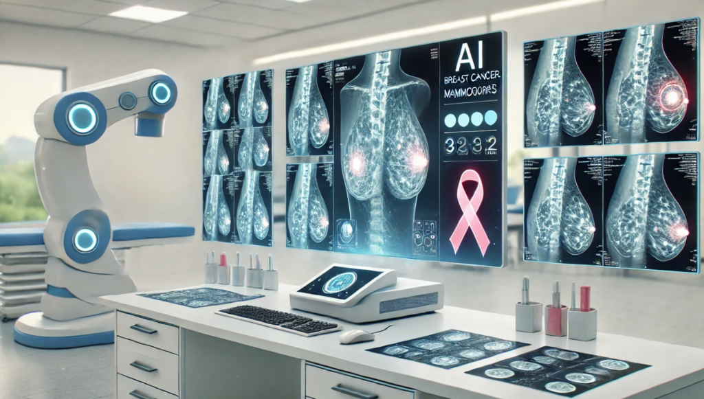
One of the most widely adopted computer-aided diagnosis (CAD) systems is in the field of breast cancer detection, particularly through mammography and histopathological imaging. Early detection of breast cancer is crucial for improving patient outcomes. However, radiology specialists often face challenges in interpreting mammograms due to the subtlety of certain abnormalities, which can lead to missed diagnoses.
CAD provides an additional layer of review, flagging suspicious areas that may require further evaluation. Advanced machine learning algorithms, particularly deep neural networks (DNNs), are used to identify potential lesions or masses, which are common early signs of breast cancer. A study in Computer Methods and Programs in Biomedicine [1] found that integrating CAD systems significantly improves the detection of early-stage breast cancer by increasing the accuracy of classification.
The study showed that deep learning models such as ResNet, InceptionV3Net, and ShuffleNet are highly effective at detecting and classifying breast cancer, with ResNet achieving 99.7% accuracy in binary classification. These CAD systems help reduce false negatives and assist in differential diagnosis, aiding radiologists in distinguishing between benign and malignant findings. While CAD should not replace the expertise of radiologists, it acts as an essential tool to improve diagnostic accuracy and patient outcomes.
CAD for Lung Nodule Detection

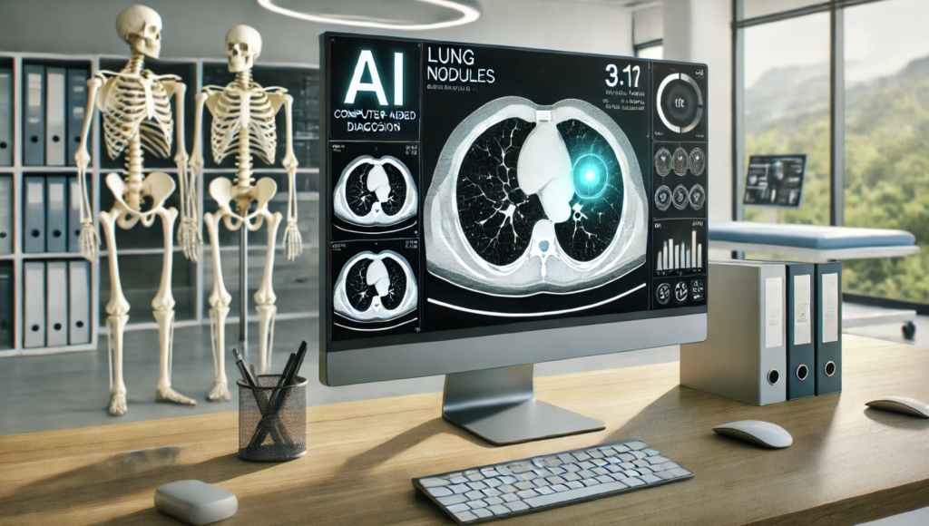
Lung nodules, small abnormal growths in the lungs, are often early indicators of lung cancer, making their detection critical. Detecting these nodules in chest CT scans can be challenging due to their size, location, and the complexity of CT images. This is where computer-aided detection systems come into play.
CAD of Liver Lesions

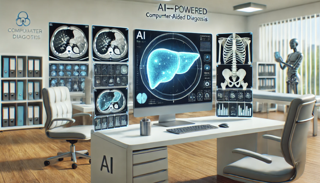
In the detection and diagnosis of liver lesions, including hepatocellular carcinoma (HCC), CAD plays an essential role. Identifying liver lesions through CT and MRI can be challenging due to the liver’s complex structure and the varying appearance of lesions across imaging modalities.
CAD for liver lesion detection utilizes AI-driven algorithms to analyze imaging data and flag regions of interest that may indicate lesions. These systems help radiologists by improving the detection of smaller lesions that might otherwise go unnoticed in traditional diagnostic approaches. Furthermore, machine learning models are trained to distinguish between different types of lesions, such as cysts, benign tumors, or cancerous growths, enabling more accurate differential diagnosis.
A study in Computers in Biology and Medicine [3] highlighted how CAD systems reduce the rate of missed lesions, improving early detection and treatment outcomes for liver diseases. By integrating AI tools with imaging platforms, CAD enhances both the speed and accuracy of liver lesion diagnostics, supporting clinicians in making informed decisions about patient care.
Enhancing Diagnostics Innovations with CAD Technologies
The integration of CAD technologies in healthcare is enhancing diagnostics in a number of ways:
Faster Analysis: CAD technologies process images quickly, enabling healthcare providers to diagnose conditions faster and reduce patient waiting times.
Minimized Errors: With the assistance of machine learning and artificial intelligence, CAD reduces the chance of human error in analyzing complex medical images.
Integration with Existing Workflows: Computer-aided diagnosis seamlessly integrates with current imaging tools, making it easy for radiologists to adopt them without overhauling existing workflows.
Wider Applications: From the detection of breast cancer to lung nodules and liver lesions, CAD systems are improving the diagnostic imaging process across many fields of medicine.
CAD technologies continue to provide significant benefits to healthcare by improving the accuracy, speed, and efficiency of diagnostic imaging.
Consult Us for AI Integration in Healthcare
If you’re looking to integrate AI and CAD technologies into your healthcare practice, Neural Board can provide expert advisory services tailored to your needs. Contact us today to learn how we can help you enhance your diagnostic capabilities with cutting-edge AI tools.
References
- Aljuaid, Hanan, et al. “Computer-aided diagnosis for breast cancer classification using deep neural networks and transfer learning.” Computer Methods and Programs in Biomedicine 223 (2022): 106951.
- Ziyad, Shabana R., Venkatachalam Radha, and Thavavel Vayyapuri. “Overview of computer aided detection and computer aided diagnosis systems for lung nodule detection in computed tomography.” Current Medical Imaging 16.1 (2020): 16-26.
- Nayantara, P. Vaidehi, et al. “Computer-aided diagnosis of liver lesions using CT images: A systematic review.” Computers in Biology and Medicine 127 (2020): 104035.
Q: What role does artificial intelligence play in computer-aided diagnosis in medicine?
A: Artificial intelligence enhances computer-aided diagnosis (CAD) systems by utilizing algorithms, such as artificial neural networks, to analyze medical images and improve diagnostic accuracy, particularly in fields like radiology.
Q: How has imaging technology evolved in the context of computer-aided diagnosis?
A: Imaging technology has significantly advanced, leading to improved resolution and clarity in medical images, which aids CAD systems in more accurately identifying conditions such as lung cancer and nodules in CT scans.
Q: What is the significance of a CAD system in mammography?
A: A CAD system in mammography assists radiologists by highlighting potential areas of concern in breast images, thus improving the early diagnosis of breast cancer and enhancing the overall accuracy of mammography screenings.
Q: Can you explain the historical review of computer-aided diagnosis in medical imaging?
A: The historical review of computer-aided diagnosis in medical imaging outlines the evolution of CAD technologies, starting from early detection methods to the integration of sophisticated computer algorithms and artificial intelligence, which have dramatically improved diagnostic capabilities.
Q: What are the current status and future potential of CAD systems in radiology?
A: The current status of CAD systems in radiology shows promising advancements in diagnostic accuracy through improved algorithms and imaging techniques, with future potential focusing on increased integration of AI and machine learning for real-time decision support in clinical use.
Q: How do CAD algorithms assist in the diagnosis of lung cancer?
A: CAD algorithms assist in the diagnosis of lung cancer by utilizing computer vision techniques to analyze imaging data, such as CT scans, to identify early signs of disease, including the detection of pulmonary nodules.
Q: What are the aspects of CAD that are crucial for clinical trials?
A: Crucial aspects of CAD for clinical trials include the validation of CADx systems, assessment of diagnostic performance, and the ability to integrate CAD findings into clinical workflows, which helps in evaluating the effectiveness of new diagnostic techniques.
Q: How does computerized detection of lung nodules contribute to modern radiology?
A: Computerized detection of lung nodules enhances modern radiology by providing radiologists with automated tools that improve the speed and accuracy of diagnoses, ultimately leading to better patient outcomes in lung disease management.
Q: What are the challenges faced by computer-aided diagnosis schemes in medical imaging?
A: Challenges faced by computer-aided diagnosis schemes include variability in image quality, the need for large datasets for training algorithms, integration into existing clinical workflows, and ensuring the reliability and interpretability of CAD outputs.
Q: What is the importance of clinical trials in validating CAD systems?
A: Clinical trials are essential for validating CAD systems as they provide evidence of the system’s diagnostic accuracy and effectiveness in real-world clinical settings, ensuring that the technology can be reliably used to support healthcare professionals in making informed decisions.

