Three-dimensional imaging has transformed diagnostic medicine, offering healthcare professionals a clearer and more detailed understanding of anatomy and pathology. Unlike traditional 2D data, 3D imaging technologies provide enhanced depth perception, allowing radiologists and surgeons to make more precise clinical decisions. The development of 3D imaging has expanded its applications across fields such as diagnostic imaging, functional magnetic resonance imaging (MRI), computed tomography (CT), and ultrasound, improving both image quality and patient outcomes.
Understanding 3D Imaging in Medicine
The introduction of 3D imaging has revolutionized the department of radiology, providing specialists with more accurate tools for medical applications. By integrating 3D data sets, clinicians can assess complex structures with greater clarity. This approach is particularly valuable for preoperative planning, trauma assessments, and monitoring chronic conditions, as it provides a more comprehensive view of the patient’s condition.
Why 3D Imaging Is Essential in Healthcare
The benefits of 3D imaging extend beyond visualization. It plays a vital role in clinical applications, making diagnostics more efficient and treatments more precise.
Key advantages of 3D imaging in medicine:
More accurate diagnoses: 3D analysis provides detailed anatomical insights, reducing misdiagnoses and improving disease detection.
Better surgical planning: The creation of 3D models allows surgeons to practice procedures before performing them on patients, leading to safer interventions.
Less invasive procedures: Techniques such as 3D volume rendering and cross-sectional images help reduce the need for exploratory surgery.
Improved communication with patients and referring physicians: 3D views help both healthcare professionals and patients understand conditions more clearly, leading to better-informed decisions.
Faster and easier-to-read imaging: 3D imaging allows for real-time adjustments and interactive exploration, making it more effective for time-sensitive diagnoses.
3D CT/MRI for Precise Diagnosis of Spinal Cord Injuries and Disc Herniation

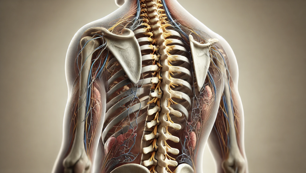
A study by Nagamatsu et al. (2022) [1] explores 3D CT/MRI fusion imaging for evaluating lumbar disc herniation and Kambin’s triangle, a critical space used in minimally invasive spine surgery. The researchers aimed to assess whether this advanced imaging approach could improve surgical planning and patient safety.
The study analyzed 3D data sets from 23 patients undergoing spinal surgery. By combining CT and MRI, researchers generated a 3D model of nerve roots and bony structures, enabling more accurate measurements of Kambin’s triangle and 3D surface angulation. Results revealed that the space available for inserting surgical instruments varied by spinal level, with the L4/5 region offering the most accessible pathway for minimally invasive procedures. However, the study also highlighted the risk of nerve compression, as surgical tools could safely pass through the triangle in only 37% of cases.
This research demonstrates that accurate 3D imaging improves diagnostic imaging of spinal structures, enhancing surgical safety. The ability to analyze 3D nerve pathways and bone alignment helps referring physicians make better preoperative assessments, reducing complications and improving patient outcomes.
3D CT Scans for Cardiovascular Disease Diagnosis and Stent Placement

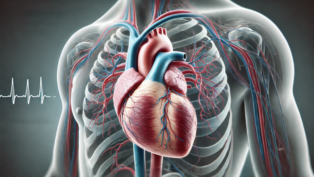
Suntharos et al. (2017) [2] investigated the use of real-time 3D CT/MRI image registration to assist in interventional procedures for congenital heart disease and acquired pulmonary vein stenosis. Traditional catheterization techniques rely heavily on fluoroscopy, which exposes patients to radiation and contrast agents. This study tested whether integrating 3D imaging techniques with live fluoroscopy could enhance procedural accuracy while minimizing radiation exposure.
Thirty patients undergoing cardiac interventions were examined using a biplane C-arm CT system, which allowed 3D volume rendering of heart structures. After registration, cross-sectional images from 3D imaging technologies were overlaid onto fluoroscopic guidance, improving catheter navigation. The study found that 64% of cases had a registration accuracy within 0–2 mm, significantly aiding images of the heart during procedures.
Although some discrepancies occurred due to motion artifacts, the findings confirm that 3D CT scans improve procedural success rates and reduce contrast-related complications. This approach enables healthcare professionals to better plan interventions such as stent placement, valve replacement, and complex congenital defect repairs, improving both benefits to patients and benefits to physicians.
3D Ultrasound for Prenatal Screening and Fetal Anomaly Detection

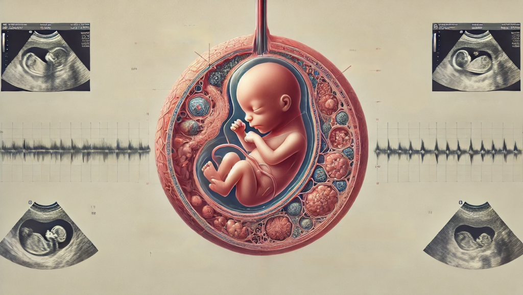
A review by Yousefpour Shahrivar et al. (2023) [3] examines machine learning-based approaches for improving fetal anomaly detection using 3D ultrasound. Traditional prenatal screening heavily depend on clinician expertise, making diagnostic accuracy variable. This study explores how AI models such as convolutional neural networks (CNNs) and U-Net segmentation can enhance fetal assessment.
The research highlights how 3D information from ultrasound scans can be analyzed using deep learning techniques to detect congenital abnormalities, including neural tube defects and heart malformations. The review discusses GANs (generative adversarial networks), which help create synthetic 3D data to train AI models, reducing biases caused by small sample sizes.
Challenges remain, particularly regarding dataset availability and clinical integration. However, the findings confirm that AI-enhanced 3D ultrasound can improve early anomaly detection, offering a less invasive alternative to traditional screening methods. Referring physicians benefit from increased diagnostic confidence, while patients receive more accurate prenatal assessments.
The Future of 3D Imaging in Healthcare
The technology has become a cornerstone of modern medicine, with 3D imaging techniques advancing rapidly. Different research institutions are driving innovation in 3D analysis, particularly in brain imaging, functional MRI, and robotic-assisted surgery.
Future 3D imaging technologies will integrate AI and real-time rendering, allowing clinicians to manipulate 3D structures interactively. Images of bones, images of the teeth, and vascular networks will become clearer and more precise, aiding both diagnostic imaging and surgical applications. AI will continue refining high-resolution images, enhancing both efficiency and accuracy.
Contact Us for AI-Powered 3D Imaging Solutions
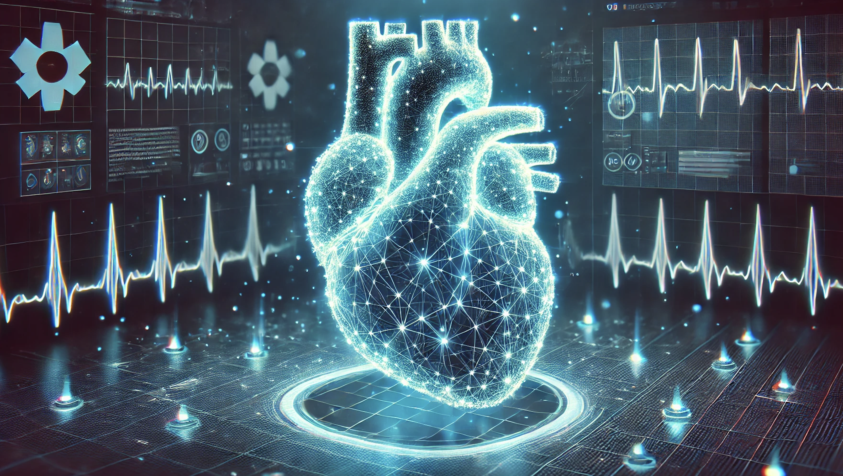
AI-driven 3D imaging is shaping the future of medical diagnostics. Our expertise in advanced imaging solutions can help healthcare professionals optimize workflows, improve accuracy, and integrate AI into existing imaging modalities. Contact us to explore custom AI in healthcare solutions for your medical practice.
References
Nagamatsu, Masakazu, et al. “Usefulness of 3d ct/mri fusion imaging for the evaluation of lumbar disc herniation and kambin’s triangle.” Diagnostics 12.4 (2022): 956.
Suntharos, Patcharapong, et al. “Real-time three dimensional CT and MRI to guide interventions for congenital heart disease and acquired pulmonary vein stenosis.” The International Journal of Cardiovascular Imaging 33 (2017): 1619-1626.
Yousefpour Shahrivar, Ramin, Fatemeh Karami, and Ebrahim Karami. “Enhancing fetal anomaly detection in ultrasonography images: a review of machine learning-based approaches.” Biomimetics 8.7 (2023): 519.

